

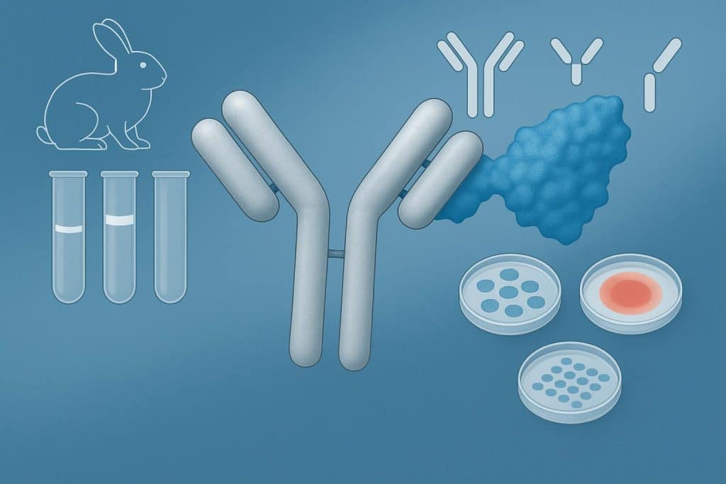
Rabbit immunoglobulin G is one of the most relied-upon antibody classes in biomedical research. Its high binding affinity across a broad antigen range, paired with a well-documented structure, makes it a consistent performer across experimental workflows. A mismatched preparation can degrade assay sensitivity, inflate background signal, or produce data that simply cannot be reproduced with confidence.
Purity Grades Explained
Suppliers offering rabbit IgG for research typically organize their catalogs by grade and application type, making it easier to narrow options based on what the assay actually requires rather than defaulting to a general-purpose preparation. Rabbit IgG is not a single standardized product—the grade reflects how thoroughly the antibody has been separated from other proteins present in serum or biological matrices, and that degree of refinement has direct consequences for assay performance.
Whole Serum vs. Purified IgG
Whole serum is a complex mixture. Alongside IgG, it contains albumin, complement components, and a range of other immunoglobulins. For basic blocking steps, it can be serviceable, but the protein variability introduces inconsistency that becomes difficult to manage in quantitative work. Purified IgG preparations strip away the bulk of those contaminants, delivering a more defined reagent with predictable protein content per unit volume.
Affinity-Purified Preparations
Protein A and Protein G affinity resins capture IgG selectively through interactions with the Fc region. This approach routinely yields preparations exceeding 95 percent IgG purity by protein content. Affinity-purified rabbit IgG is the practical standard for assays where low background matters, including enzyme-linked immunosorbent assays and multi-parameter flow cytometry panels.
High-Purity Grades for Sensitive Assays
Certain workflows set a higher bar. Preparations further refined through ion-exchange chromatography reduce residual non-IgG protein to levels that more standard purification methods leave behind. Surface plasmon resonance, biolayer interferometry, and proteomics applications using mass spectrometry fall into this category. For these assays, even minor contaminants can obscure binding kinetics or introduce confounding signals.
Common Formats and Their Uses
Purity addresses what is in the preparation. Format addresses how the antibody behaves once it is in the assay.
Intact IgG
The full antibody retains both antigen-binding fragments and the crystallizable fragment. This bivalent structure supports secondary antibody recognition through the Fc region, making intact IgG the default choice for Western blotting, immunohistochemistry, and indirect immunofluorescence. Its compatibility with standard detection systems keeps it versatile across these platforms.
F(ab’)₂ and Fab Fragments
Tissues with high endogenous Fc receptor activity present a specific challenge. Intact IgG binds non-specifically through its Fc region in those environments, creating background that obscures genuine signal. F(ab’)₂ fragments retain bivalent antigen binding while removing the Fc region entirely. Fab fragments take this process further, offering monovalent binding for applications requiring even tighter control. Both are generated through enzymatic digestion and are particularly valuable in multiplex immunofluorescence panels where staining specificity cannot be compromised.
Biotinylated and Conjugated Forms
Pre-conjugated rabbit IgG reduces protocol complexity without sacrificing detection performance. Biotinylated preparations integrate directly into streptavidin-based amplification systems. Enzyme-conjugated forms, most commonly horseradish peroxidase-linked, remain a practical staple in blotting and plate-based detection assays. Fewer incubation steps mean fewer variables, and that translates to more reproducible results across runs.
Matching Grade and Format to Application
The decision comes down to mapping reagent specifications to what the assay actually demands.
Standard affinity-purified intact IgG handles routine Western blotting and immunoprecipitation without overcomplicating procurement. Flow cytometry panels benefit from low-endotoxin, high-purity preparations, since cellular health and non-specific binding are both sensitive to contaminant levels. Structural and biophysical studies require the strictest purity specifications, with endotoxin quantified and aggregate content assessed by size exclusion chromatography before use.
Tissue-based assays may call for F(ab’)2 fragments depending on receptor expression in the target tissue and the secondary detection system in play. Multiplex panels require format coordination across every reagent in the stack, not just the primary antibody.
Conclusion
Rabbit IgG antibodies are dependable across a wide range of research applications, but that dependability is conditional. It depends on selecting a preparation whose purity grade and format align with what the assay requires. Purity shapes background levels and signal reliability; format governs binding behavior and compatibility with detection systems. Evaluating both criteria before procurement is one of the more straightforward ways to reduce variability and avoid the kind of troubleshooting that costs time without improving the underlying science.